

HERE’S AN OFFER most people can easily refuse: Donate part of your brain to science — while you’re still alive.
But in the Seattle area, about 50 people every year say, “Sure.”
Gary Williams is one of them.
The former logger and Army veteran suffers from epilepsy so severe he was forced to give up the job he loved as a tattoo artist. The electrical storms in his brain robbed him of a near-photographic memory. He can’t even ride his mountain bike anymore for fear the exertion will bring on an attack.
So on the morning of his 48th birthday, Williams is lying in a bed at Harborview Medical Center, waiting to be wheeled into the operating room. Bearded and heavily inked with designs that range from Sasquatch and Mount Rainier to a pot-smoking gypsy, he also sports a small checkmark on his left temple. It confirms the spot where neurosurgeons will soon open his skull and attempt to cut out the part of his brain responsible for seizures that wrack his body and leave him limp and shattered for days at a time.
In order to reach that faulty patch of brain, Williams’ surgeon will first have to snip out a marble-sized piece of healthy cerebral cortex — the wrinkled, outermost layer of the brain, where our higher cognitive abilities reside. At most hospitals, the bits of normal tissue excised during surgeries for epilepsy or brain tumors are burned as medical waste. But to scientists in Seattle, they’re a treasure trove — an astonishingly rare opportunity to peer into the workings of the living human brain.
By rushing the tissue from the OR to laboratories in South Lake Union, researchers at the Allen Institute for Brain Science can study the cells while they’re still alive and connected to each other, crackling with the electrical impulses that are the currency of thoughts, memories and perception. Many samples remain viable for three days. With special handling and treatment, some of the tiny slices will continue to function for a month or more.
If you’ve watched many horror movies, the notion of scientists zapping human brain tissue in the lab might conjure images of Dr. Frankenstein screaming, “It’s ALIVE!” But these bits of tissue lack anything approaching sentience, and the Seattle work has more noble goals. The loftiest is a better understanding of how billions of neurons collectively give rise to human consciousness. Studying live human brain tissue is the only way to learn how those neurons actually function within the body’s most complicated organ, the researchers say. And because decades of mouse studies have yet to yield cures for Alzheimer’s, autism, epilepsy and a host of other vexing disorders, many experts argue that live human brain tissue might offer a more effective way to figure out what goes awry in diseases and how to fix it.
“This is very cutting-edge stuff,” says neuroscientist Jonathan Ting, a leader of the project. “We don’t even know the limits of what we can do or the questions we can ask with these techniques.”
None of it would be possible without people like Williams, who’s happy to see his surgical leftovers put to good use.
“Why wouldn’t I want to help other people that have the same problems as me?” he asks, shrugging.
FOR A MAN about to have a 3-inch hole cut in his head, Williams is remarkably calm.
“I’m a combat veteran,” he says, as machines beep and monitor his vital signs. “I’ve been through worse.”
During the operation, UW Medicine neurosurgeon Dr. Jeffrey Ojemann will do his best to avoid critical areas of Williams’ brain. But the procedure carries risks, including memory and speech impairment. Williams is betting on the upside: that he will have fewer seizures and a clearer head.
“I’m being reborn,” he says, with a grin.
Ninety minutes later, Williams is out cold and covered with blue surgical drapes. Only the left side of his cranium is visible. After bone is cut and the tough, protective dura peeled back, Williams’ brain is exposed, glistening and pulsing gently. Ojemann slides a strip of electrodes into the area where he suspects the epilepsy originates. EEG tracings race across a screen, some jumbled and staccato.
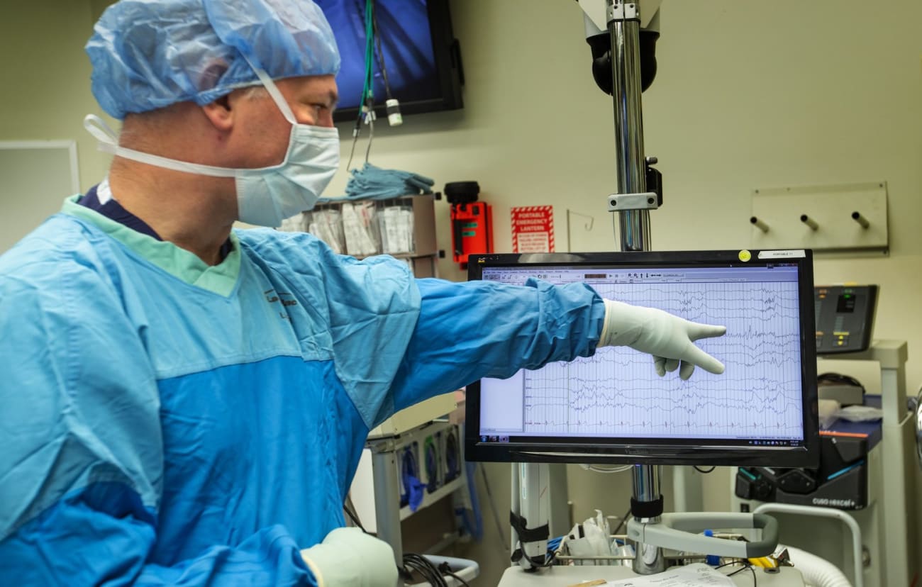
UW Medicine neurosurgeon Dr. Jeffrey G. Ojemann points to abnormal brainwave patterns detected after he placed electrodes on the surface of Gary Williams’ brain to pinpoint the region causing epileptic seizures.
“That’s really abnormal,” Ojemann says, pointing to a pattern of frenetic peaks. “It’s like a mini-seizure.”
Ojemann was born to the brain business. His father and brother are neurosurgeons, as was his late uncle; his mother is a neurologist who specializes in epilepsy. When he’s not operating, Ojemann analyzes brainwave recordings to find better treatments for stroke and epilepsy. He was eager to collaborate when the Allen Institute started its human brain tissue initiative five years ago.
“I hope this helps us treat epilepsy and helps us develop new therapies,” he says. “I think the sky’s the limit on what they can learn.”
Once he’s pinpointed the troublesome portion of Williams’ brain, Ojemann is ready to begin cutting. Peering through a surgical microscope, he teases away a disc of tissue from the outermost layer of the temporal lobe, a part of the cerebral cortex involved in memory, speech and comprehension. He places the pinkish blob in a beaker filled with a frozen slurry of artificial cerebrospinal fluid.
It’s just the beginning of Williams’ surgery, which will continue for another two hours. But the race is on to get that morsel of brain into the lab.